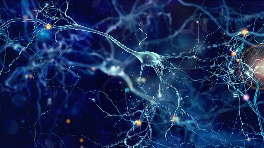
In a latest research printed in Cell Stem Cell, researchers produced three-dimensional (3D) bioprinted human mind tissues, permitting for the creation of functioning neural networks that might simulate community exercise in regular and pathological conditions.

Background
Understanding neural networks within the human mind is important for understanding mind well being and illness. Nevertheless, animal-based fashions can not successfully reproduce the human mind’s high-order information processing as a result of variations in cell composition, neural networks, and synaptic integration. 3D bioprinting offers a extra correct methodology for creating human mind tissues by bodily repositioning hydrogels and stay cells inside a physiologically difficult cytoarchitecture. Nevertheless, bioprinting smooth tissues, such because the mind, is of concern since smooth biomaterials can not maintain intricate 3D architectures or inflexible gels.
In regards to the research
Within the current research, researchers developed a 3D bioprinting platform to fabricate tissues with outlined human mind cell sorts in any desired dimension.
The crew aimed to construct layered neural tissues, together with neural progenitor cells (NPCs) that generate connections inside and between the mind layers, sustaining the construction intact. They created a bioink for printing. They used fibrin gel to print the tissues. Strategies for bioprinting embody extrusion-based, laser-based, and droplet-based strategies. The extrusion three-dimensional bioprinting approach deposited gel in layers to simulate mind buildings corresponding to human cortex laminations.
The researchers chosen a 50 mm thickness for each layer and constructed multi-layered tissues by inserting the layers in a horizontal association adjoining to at least one one other. They designed 3D-printed mind tissues to be comparatively skinny however purposeful and multi-layered, with established cell compositions and fascinating dimensions, and simply maintained and examined in a normal laboratory setting.
The researchers decided that 2.50 mg per mL fibrinogen and 0.50 to 1.0 U of thrombin had been optimum concentrations for hydrogel formation, leading to a gelation period of 145 seconds, which allowed for 24-well plate printing. After six hours, most (85%) of the cells had been viable and survived for seven days. The crew created medial ganglionic eminence (MGE)-derived gamma-aminobutyric acid (GABA) and cortical (glutamate) progenitors from inexperienced fluorescent protein-expressing (GFP+) and GFP– human pluripotent stem cells (hPSCs) to research whether or not GABAergic interneurons and glutamatergic neurons type synaptic connections when inserted into printed tissues. Earlier than printing, they mixed the 2 progenitor populations in a 1:4 ratio to match the ratio of interneurons to cortical projection neurons within the cerebral cortex.
The researchers recorded electrophysiological information from tissues printed with GFP+ glutamatergic cortical progenitors, noncolored MGE GABAergic progenitors, and hPSC-derived astrocyte progenitors integrated into glutamate neurons and GABA interneurons. The printed tissue was immunostained with an axonal marker, SMI312. They studied Alexander illness (AxD), a neurodegenerative illness brought on by GFAP gene abnormalities, to research pathogenic mechanisms. They used stay imaging of glutamate uptake by glutamate-sensitive fluorescent reporters (iGluSnFR) to research neuron-astrocyte interactions and neuron-glial connections in AxD.
Outcomes
The printed neuronal progenitors developed into neurons inside weeks, forming purposeful neural networks inside and throughout tissue layers. Printed astrocyte progenitors matured into astrocytes with advanced processes to operate in neuron-astrocyte networks. Typical tradition strategies might retain the 3D mind tissues, making them simpler to research in physiological and pathological settings. Cell viability declined with rising concentrations of thrombin at 2.50 mg/mL fibrinogen concentrations however remained unaltered at a continuing focus of 0.50 U fibrinogen, and cells aggregated at elevated fibrinogen ranges.
The bioprinted neural cells matured and retained tissue type, with GFP-expressing cells in a single band reworking into microtubule-associated protein 2 (MAP2+) neurons every week after printing. The printed tissue maintained a secure configuration the place neural progenitors multiplied and constructed neural networks. The neuronal subtypes established purposeful networks throughout the bioprinted tissues, with hPSC-derived MGE cells expressing NK2 homeobox 1 (NKX2.1) and GABA and cortical progenitors constructive for forkhead-box G1 (FOXG1) and paired field 6 (PAX6). The bioprinted neural tissue constructions promote the expansion of cortical glutamatergic neurons and GABAergic interneurons.
The researchers utilized a high-concentration potassium chloride resolution to print tissues containing neurons and astrocytes, demonstrating purposeful connections. The astrocytes expressed glutamate transporter 1 (GLT-1), indicating maturation. The printed cortical and striatal neuronal bands remained intact 15 days after printing, and GFP and mCherry neurites developed in the direction of one another. The printed human mind tissues may replicate diseased processes, with AxD astrocytes exhibiting intracellular GFAP aggregation. By 30 days, MAP2+ neurons and GFAP+ astrocytes exhibited advanced morphology and synapsin expression.
Conclusion
General, the research findings demonstrated the power of 3D printing to generate functioning mind tissues for simulating community exercise in regular and pathological settings. The bioink-created tissues set up purposeful synaptic connections between neuronal subtypes and neuron-astrocyte networks in two to 5 weeks. The 3D platform offers an outlined surroundings for finding out human mind networks in wholesome and pathological settings; nonetheless, it has limitations, such because the softness of the gel and the 50mm thickness of the printed tissues.
Supply hyperlink


Excellent write-up
Insightful piece
Your posts always provide me with a new perspective and encourage me to look at things differently Thank you for broadening my horizons
Insightful piece