Utilizing intravascular imaging to information stent implantation throughout percutaneous coronary intervention (PCI) in coronary heart illness sufferers considerably improves survival and reduces antagonistic cardiovascular occasions in comparison with angiography-guided PCI alone, essentially the most generally used methodology.
These are the outcomes from the biggest and most complete scientific research of its variety evaluating two sorts of intravascular imaging strategies (intravascular ultrasound, or IVUS, and optical coherence tomography, or OCT) with angiography-guided PCI. The research, revealed Wednesday, February 21, in The Lancet, is the primary to indicate that these two strategies of high-resolution imaging can cut back all-cause demise, coronary heart assaults, stent thrombosis, and the necessity for revascularization.
Our research, representing a synthesis of all early and up to date scientific research, has proven for the primary time that the routine use of intravascular imaging steering improves survival and enhances all points of the protection and effectiveness of coronary stenting, even with glorious up to date drug-eluting stents.”
Gregg W. Stone, MD, first creator
Dr. Stone is Director of Educational Affairs for the Mount Sinai Well being System, and Professor of Drugs (Cardiology), and Inhabitants Well being Science and Coverage, on the Icahn College of Drugs at Mount Sinai.
“Prior research had proven advantages of intravascular imaging, however by no means to this extent,” Dr. Stone provides. “The addition of 4 latest trials through which 7,224 sufferers have been enrolled now exhibits that intravascular imaging reduces all-cause demise and all coronary heart assaults throughout the big selection of sufferers who endure stent remedy. As such, the routine use of intravascular imaging to information stent implantation is without doubt one of the best therapies we now have to enhance the prognosis of sufferers with coronary artery illness.”
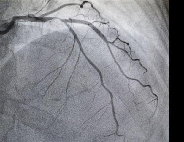
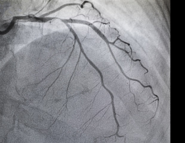
Sufferers with coronary artery disease-;plaque buildup contained in the arteries that results in chest ache, shortness of breath, and coronary heart attack-;typically endure PCI, a non-surgical process through which interventional cardiologists use a catheter to position stents within the blocked coronary arteries to revive blood circulate. Interventional cardiologists mostly use angiography to information PCI, which entails a particular dye (distinction materials) and X-rays to see how blood flows by way of the guts arteries to spotlight any blockages.
Angiography has limitations, nonetheless, making it tough to find out the true artery dimension and the make-up of the plaque, and is suboptimal in figuring out whether or not the stent is totally expanded post-PCI and in detecting different situations that have an effect on the early and late outcomes of the process. Intravascular ultrasound was launched greater than 30 years in the past to offer a extra correct and particular image of the coronary arteries. Despite the fact that research have proven that IVUS-guided PCI is superior to angiography-guided PCI and reduces cardiovascular occasions, it is just utilized in roughly 15 to twenty % of PCI instances in the USA, because the pictures could also be tough to interpret and the process isn’t totally reimbursed.
Optical coherence tomography makes use of gentle as a substitute of sound to create pictures of the blockages. OCT pictures are a lot larger in decision, extra correct, and extra detailed in comparison with IVUS, and simpler to interpret. Nonetheless, as a more recent approach, OCT is utilized in solely 3 % of PCI instances, partly due to a scarcity of research data-;a limitation this new research has addressed.
Of their research, the researchers analyzed knowledge from 15,964 sufferers present process PCI from 22 trials in a whole bunch of facilities from the USA, Europe, Asia, and elsewhere between March 2010 and August 2023. Sufferers underwent both angiography-guided PCI or intravascular imaging-guided PCI utilizing both IVUS or OCT. Throughout follow-up starting from 6-60 months with a imply of two years, sufferers who acquired intravascular imaging steering skilled a 25 % discount in all-cause demise, 45 % discount in cardiac demise, 17 % discount in all myocardial infarctions, and 48 % discount in stent thrombosis in contrast with angiography steering. The research additionally discovered that intravascular imaging lowered goal vessel myocardial infarction by 18 % and goal lesion revascularization by 28 %. The outcomes have been related for OCT-guided and IVUS-guided PCI.
“With these outcomes, we now must shift from performing extra research to find out whether or not intravascular imaging is useful, to rising efforts to beat the remaining impediments to the routine use of OCT and IVUS, together with higher coaching of physicians and workers and rising reimbursement,” Dr. Stone stated. “On this regard, we now have higher ‘laborious proof’ that intravascular imaging steering of PCI procedures makes a larger affect to bettering our sufferers’ lives than different routine therapies that are extra broadly used and reimbursed.”
Supply:
Journal reference:
Stone, G. W., et al. (2024). Intravascular imaging-guided coronary drug-eluting stent implantation: an up to date community meta-analysis. The Lancet. doi.org/10.1016/S0140-6736(23)02454-6.
Supply hyperlink


:max_bytes(150000):strip_icc()/700A4310-2a4ddccea527498e9f69834d730d4d55.jpg)


