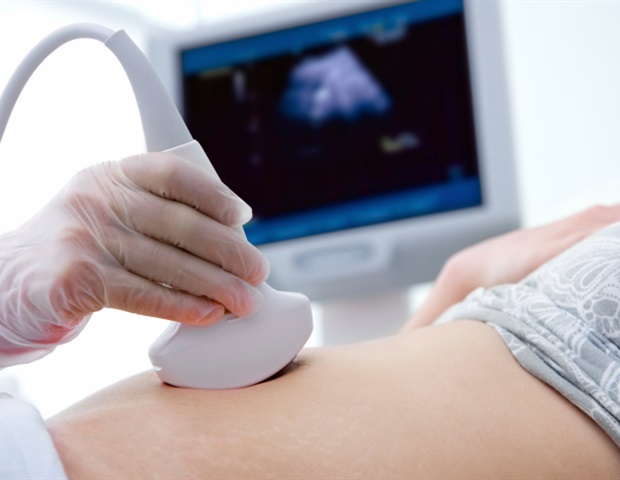
Synthetic intelligence can spot COVID-19 in lung ultrasound pictures very like facial recognition software program can spot a face in a crowd, new analysis reveals.
The findings increase AI-driven medical diagnostics and convey well being care professionals nearer to with the ability to rapidly diagnose sufferers with COVID-19 and different pulmonary illnesses with algorithms that comb by ultrasound pictures to establish indicators of illness.
The findings, newly revealed in Communications Medication, culminate an effort that began early within the pandemic when clinicians wanted instruments to quickly assess legions of sufferers in overwhelmed emergency rooms.
We developed this automated detection device to assist medical doctors in emergency settings with excessive caseloads of sufferers who should be identified rapidly and precisely, comparable to within the earlier phases of the pandemic. Probably, we need to have wi-fi units that sufferers can use at dwelling to observe development of COVID-19, too.”
Muyinatu Bell, senior writer, the John C. Malone Affiliate Professor of Electrical and Laptop Engineering, Biomedical Engineering, and Laptop Science at Johns Hopkins College
The device additionally holds potential for growing wearables that monitor such diseases as congestive coronary heart failure, which might result in fluid overload in sufferers’ lungs, not in contrast to COVID-19, mentioned co-author Tiffany Fong, an assistant professor of emergency medication at Johns Hopkins Medication.
“What we’re doing right here with AI instruments is the subsequent massive frontier for level of care,” Fong mentioned. “An excellent use case could be wearable ultrasound patches that monitor fluid buildup and let sufferers know after they want a drugs adjustment or when they should see a health care provider.”
The AI analyzes ultrasound lung pictures to identify options often called B-lines, which seem as brilliant, vertical abnormalities and point out irritation in sufferers with pulmonary problems. It combines computer-generated pictures with actual ultrasounds of sufferers -; together with some who sought care at Johns Hopkins.
“We needed to mannequin the physics of ultrasound and acoustic wave propagation properly sufficient so as to get plausible simulated pictures,” Bell mentioned. “Then we needed to take it a step additional to coach our pc fashions to make use of these simulated information to reliably interpret actual scans from sufferers with affected lungs.”
Early within the pandemic, scientists struggled to make use of synthetic intelligence to evaluate COVID-19 indicators in lung ultrasound pictures due to a scarcity of affected person information and since they have been solely starting to grasp how the illness manifests within the physique, Bell mentioned.
Her group developed software program that may be taught from a mixture of actual and simulated information after which discern abnormalities in ultrasound scans that point out an individual has contracted COVID-19. The device is a deep neural community, a kind of AI designed to behave just like the interconnected neurons that allow the mind to acknowledge patterns, perceive speech, and obtain different complicated duties.
“Early within the pandemic, we did not have sufficient ultrasound pictures of COVID-19 sufferers to develop and check our algorithms, and consequently our deep neural networks by no means reached peak efficiency,” mentioned first writer Lingyi Zhao, who developed the software program whereas a postdoctoral fellow in Bell’s lab and is now working at Novateur Analysis Options. “Now, we’re proving that with computer-generated datasets we nonetheless can obtain a excessive diploma of accuracy in evaluating and detecting these COVID-19 options.”
Supply:
Journal reference:
Zhao, L., et al. (2024). Detection of COVID-19 options in lung ultrasound pictures utilizing deep neural networks. Communications Medication. doi.org/10.1038/s43856-024-00463-5
Supply hyperlink


